PROJECT
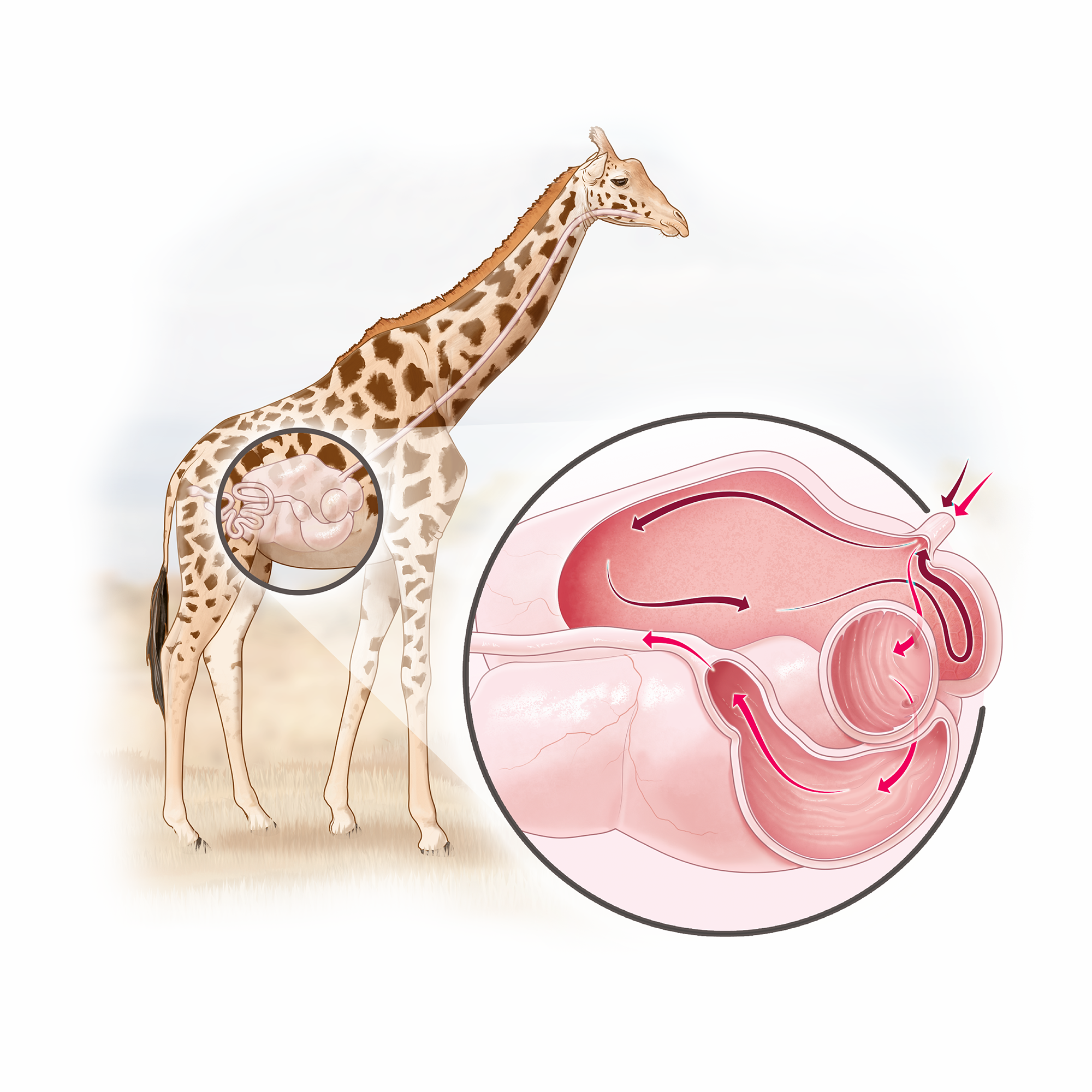
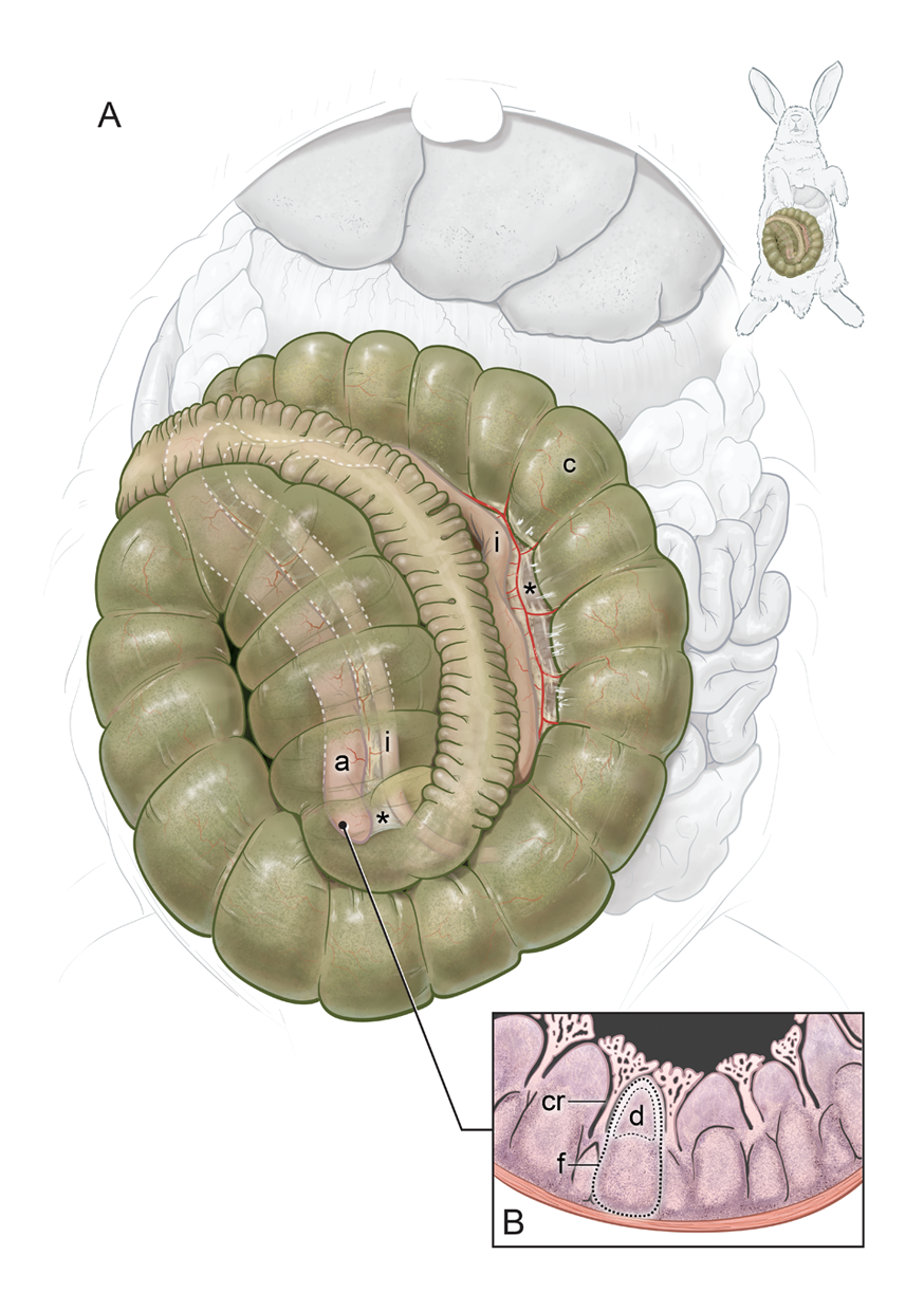
I was commissioned to create a detailed anatomical illustration depicting the elusive ileocecal fold and adjacent structures in healthy rabbits. This illustration would be included in a very complete scientific article discussing appendicitis in rabbits, to be published in the Journal of the American Veterinary Medical Association.
After accessing all the relevant structures to highlight, it was important to come up with the most informative view. The client and I decided to depict the body in a dorsal decubitus position, with all the gastrointestinal organs in their normal positions. This would make it easier for veterinarians to localize the ileocecal fold during a necropsy. Because this fold is hidden under the large cecum, we played with colors, contrast and transparency to create an accurate and informative representation.
Finally, since this was to be published in a scientific journal, we had to adhere to the figure guidelines, ensuring a smooth review and acceptance process.
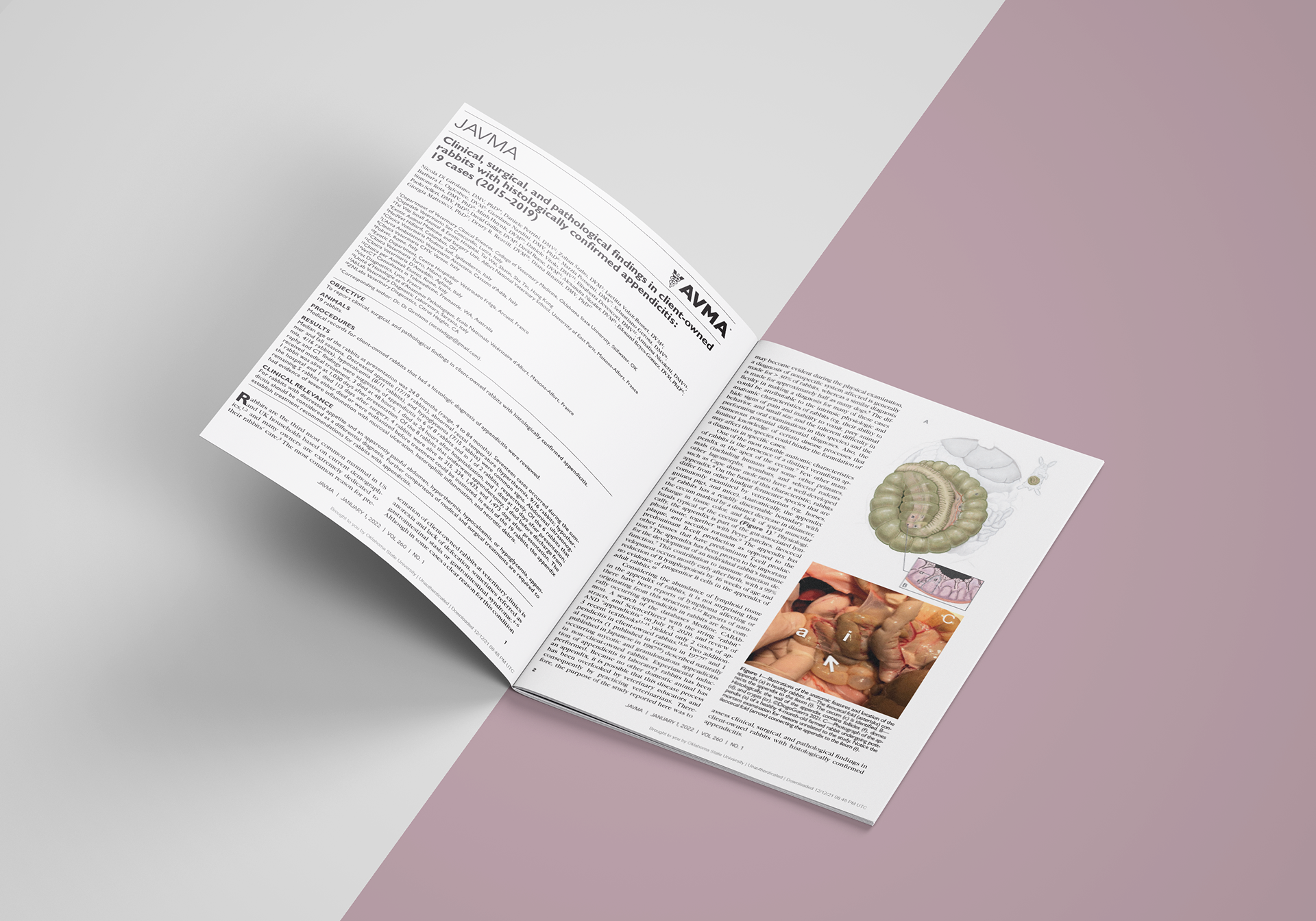
Read the full publication here.
© Diogo Guerra. 2021
CLIENT
Dpt of Vet Clinical Sciences,
College of Veterinary Medicine,
Oklahoma State University, US
TOOLS
Adobe Illustrator®
Adobe Photoshop®
TOPICS
#Rabbit #Rabbitmedicine
#Gastroenterology
#MedicalIllustration
#Sciart

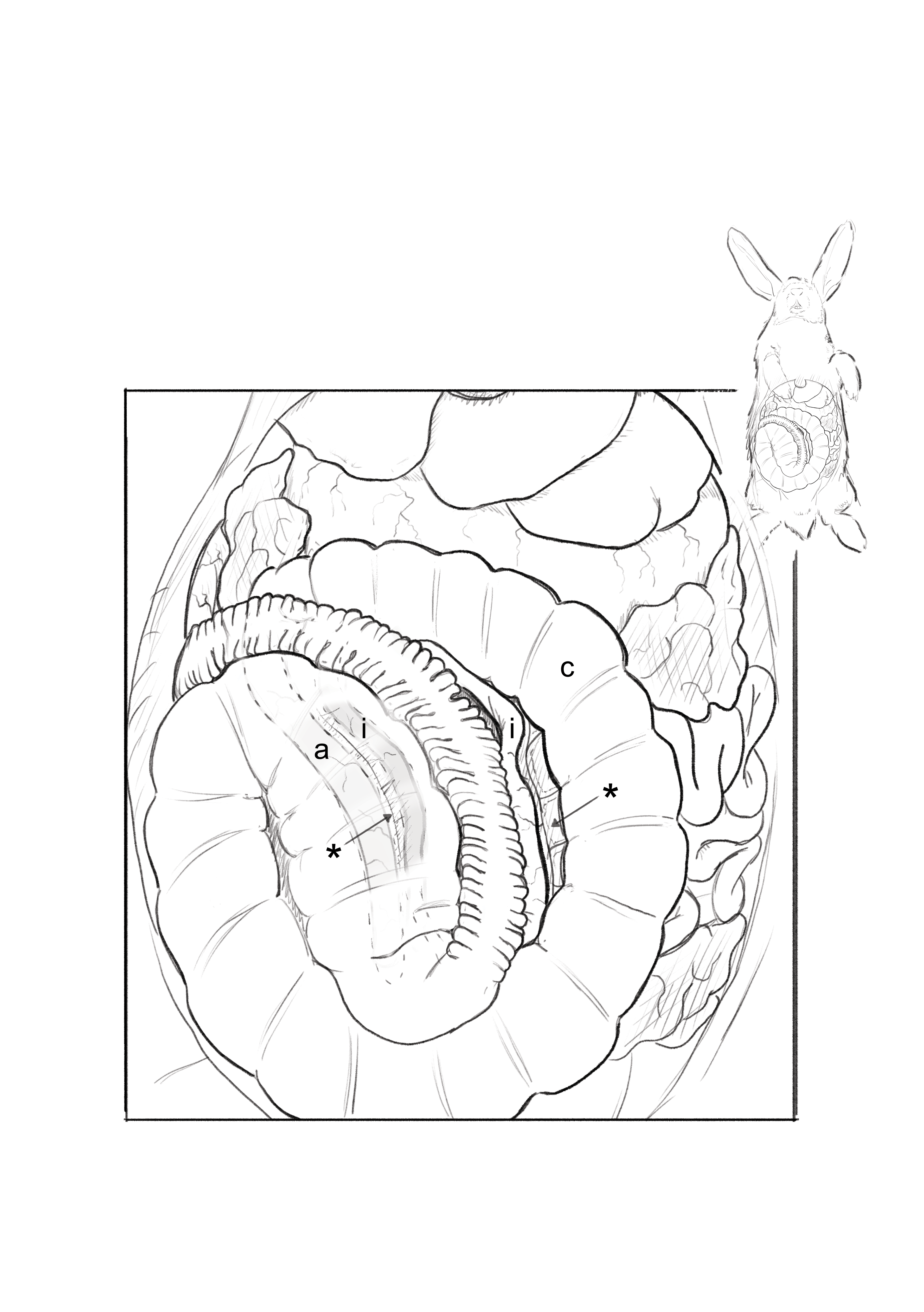
/ anatomical features and location of the appendix (a) in healthy rabbits. (A) The ileocecal fold (*) connects the appendix (a) to the ileum (i). The cecum (c) is identified. (B) Histologically, the wall of the appendix contains follicles (f), domes (d), and crypts (cr).
/ sketch in Adobe Photoshop

/ cleaned-up line art in Adobe Illustrator
/ digital painting in Adobe Photoshop
© Diogo Guerra. 2021